- 移动端

上海南方模式生物科技股份有限公司品牌商
14 年
手机商铺
- NaN
- 0
- 0
- 2
- 2
推荐产品

公司新闻/正文
Nature子刊登我公司斑马鱼和细胞技术平台与同济大学合作研究成果
1127 人阅读发布时间:2016-05-20 11:05
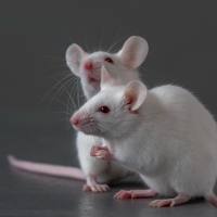
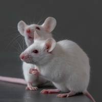
我公司斑马鱼和细胞技术平台与同济大学合作,开展了关于抗癌药物协同作用的研究。
利用细胞与斑马鱼模型对16种小分子化学药物及其多种药物组合进行体内外筛选评估(含毒理学研究和药效学研究),最终确认了两种候选药物对乳腺癌治疗具有协同效应,非小细胞肺癌,也有类似的表现。该研究结果于2015年9月28日,在Nature著名学术子刊《Nature Communications》(影响因子:11.47)在线发表。
Combining genomic and network characteristics for extended capability in predicting synergistic drugs for cancer
Abstract
The identification of synergistic chemotherapeutic agents from a large pool of candidates is highly challenging. Here, we present a Ranking-system of Anti-Cancer Synergy (RACS) that combines features of targeting networks and transcriptomic profiles, and validate it on three types of cancer. Using data on human β-cell lymphoma from the Dialogue for Reverse Engineering Assessments and Methods consortium we show a probability concordance of 0.78 compared with 0.61 obtained with the previous best algorithm. We confirm 63.6% of our breast cancer predictions through experiment and literature, including four strong synergistic pairs. Further in vivo screening in a zebrafish MCF7 xenograft model confirms one prediction with strong synergy and low toxicity. Validation using A549 lung cancer cells shows similar results. Thus, RACS can significantly improve drug synergy prediction and markedly reduce the experimental prescreening of existing drugs for repurposing to cancer treatment, although the molecular mechanism underlying particular interactions remains unknown.

(a) Seventeen agent pairs tested by experiment on MCF7 cell line. CI for each drug pair was summarized in a heat map. Green indicates synergy (CI<0.9); dark green indicates strong synergy (CI<0.3); yellow indicates additive (0.9
全文链接:http://www.nature.com/ncomms/2015/150928/ncomms9481/full/ncomms9481.html